
Old No 20, New No 33, 7th Avenue, Ashok Nagar, Chennai-600083.
Retina And Vitreous Disorders
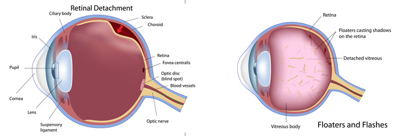
Retina is the light receptive screen of the eye (akin to the screen in the camera) that sends visual messages to the brain through the optic nerve. When the retina is lifted or pulled it's from normal position it gets detached. This interferes with the reception of light by the retina. If this is not treated immediately, this could result in permanent blindness.
Retinal detachments are caused by a host of conditions in the eye,
 Rhegmatogenous detachment – caused by retinal tears or breaks (dehiscence in the retina)
- Tractional detachment – caused by tractional bands (due to fibrous tissue growth on the surface of retina)
- Exudative detachment – caused by injury to or inflammation in the eye (when the retina is lifted by fluid accumulation behind it)
The vitreous is a gel like substance filling the eye behind the lens and in front of the retina. It is an optically clear viscous medium. The vitreous can be become opaque due to bleeding in the eye due to injury/ diabetes mellitus etc..
Retinal detachment can occur if you
- Are over 40 years of age
- Are myopic (especially high myopia)
- Had a retinal detachment in one eye
- Have a family history of retinal detachment
- Have other eye diseases like retinoschisis, uveitis, lattice degeneration, retinal breaks etc
- Had an eye injury
- Untreated vitreous hemorrhage, diabetic retinopathy etc.
Symptoms of retinal detachment/ vitreous diseases you should look out for
- Sudden appearance or increase in ``Floaters`` – black cobweb like little specks in the field of vision
- Flashes of light
- Appearance of a curtain over the field of vision
- Sudden or gradual drop in vision
Treatment of retina and vitreous diseases is done with
- Laser indirect ophthalmoscopy – for retina breaks in which the hole or break in the retina is sealed with laser
- Cryopexy – which freezes the area around the hole to reattach the retina
- Pnuemoretinopexy – where air is injected into the vitreous to push the retina to its normal position
- Scleral buckling – where a buckle is attached to outside of the eye to gently push the wall of the eye against the detached retina
- Vitrectomy – is performed with the state of the art Accurus machine in which the vitreous is removed partly or totally through a small incision in the sclera, gas is injected into the eye to push the retina to its normal position
- Over 90% of retinal detachment can be successfully treated with good visual outcome achieved when the patient presents early in the course of disease. It is vital to report to the eye specialist at the first instance of noticing floaters, flashes or curtain in the field of vision.
Treatment for retinal detachment / vitreous diseases
Vision Xperts Eye Hospital is fully equipped for the management of disorders of retina and vitreous with very experienced vitreo – retinal surgeons providing all the available treatment options. Vision Xperts Eye Hospital also has a – state – of – art machines and Operation Theater exclusively dedicated to treating vitreo – retinal conditions.
Diagnosis of retina and vitreous diseases is done with
- Indirect Ophthalmoscopy
- B – Scan ultrasound
- Optical Coherence Tomography